Eye Health & Diseases
Eye Diseases Departments of Medical Park...
Main Page
- 2
Eye Health & Diseases
- 3
Contact
Check Other Treatments
At Eye Diseases Departments of Medical Park Hospitals Group, the first step of routine eye examination is to hear the patients' complaints regarding their vision. Taking into account the complaints' characteristics, the patient's eyebrows, eyelids, and view position of the eyes are observed in the first place during routine eye examination. The patient's refractive error is then measured by means of computerized auto-refractometer and retinoscope. The visual acuity of both eyes with and without glasses is determined. The eyelashes, conjunctiva, cornea, and other elements of the anterior segment of the eye are carefully examined during biomicroscopic examination, followed by measurement of the eye pressure.
Refractive Errors

Light and images of objects are refracted by the cornea and lens of the eye to reach the spots of vision on the retina. In a normal eye, light beams coming from the outside are refracted by the cornea and lens to reach the visual cortex, which provides visual acuity.
Diverse alternatives are available to achieve sharp vision in persons with refractive errors. The options available for refractive error correction include eyeglasses, contact lenses, or treatment by excimer laser. Furthermore, the eye examination may help to diagnose retinal detachment, hypertension, brain tumor, and symptoms of diverse diseases in the body.
Cataract and its treatment (Phacoemulsification)
Cataract is the clouding of the normally clear lens of the eye. In 90% of these cases, cataract is age related, but it may be seen in all age groups including infants. The most frequently seen symptoms of cataract are painless visual loss, glare or increased sensitivity to light, paling or yellowing of colors, and deteriorated night vision.
What does retina, vitreous and macula mean?
The retina is the sensory membrane that lines the inner surface of the back of the eyeball and consists of millions of photoreceptor cells, allowing the brain to perceive images. The perceived image is transported to the brain by way of the optic nerve, thus creating vision. If we would compare the eyeball with a camera, the retina would be the sensor on which the light reflects. The midmost section of this sensor allowing for central and sharp vision is referred to as the macula, whereas the glairy fluid filling the eyeball is called vitreous. Diseases and conditions of the retina and vitreous are often interconnected.
What are the most common retinal diseases?
- Diabetic retinopathy
- Retinal break and retinal detachment
- Age-related macular degeneration (yellow spot disease)
- Macular hole
- Epiretinal membrane (macular pucker)
- Retinal vein occlusions
- Retinal artery occlusions
VITREORETINAL SURGERY
These are surgeries related to the intraocular gel-like clear structure, referred to as vitreous filling the inner chambers of the eye, and the retina. The vitreous may degenerate and result in loss of vision by various reasons (such as trauma, diabetes, uveitis, intraocular bleeding etc.). Membranes may build up in the vitreous, which may create a pulling effect on the retina, thereby giving rise to retinal break and retinal detachment. Besides, holes may occur in the macula, which is responsible for central vision, along with thin membranes on the macular surface. All these degenerations can be treated surgically. In this surgical procedure, the vitreous humor is removed by penetrating into the insides of the eye with special instruments under a microscope with the help of retinal imaging systems and replaced by special balanced solutions, bleedings are stopped, any membranes covering the retinal surface are stripped off, and attachment of the retina is restored by draining any fluids that have accumulated under the retina in cases of retinal detachment. This procedure is microsurgical, during which use is made of laser, various dyes to dye the membranes, and buffer substances like silicone or gas. These are seamless surgeries, which provide anatomic and functional restoration.
RETINOPATHY OF PREMATURITY
Retinopathy of prematurity is defined as one of the most important health problems that may affect the eyes of premature babies. The vessels in the eyes of infants continue to develop until the baby is born. This development is interrupted in premature babies and continues after birth. The high concentrations of oxygen administered to premature babies in order to keep them alive may cause abnormal development of vessels in their eyes, resulting in Retinopathy of Prematurity, shortly referred to as ROP, in the retina of babies whose vessel development is not completed in the womb. If not treated in early stages, ROP may cause blindness in both eyes. Therefore, premature babies must be examined by an ophthalmologist.
Let’s connect
We’re here to help and answer any question you might have, We look forward to hearing from you. You can use our chat box at the very bottom right corner for quick answers and furthermore questions.
Join our community
You can follow us from Instagram.
Follow us via InstagramContact Us Via Whatsapp!
You can get fast information about any treatments via Whatsapp.
Send us message
 4 April 2024
Get ready for summer!
Get ready for summer! With Hollywood Smile treatment, make a difference this summer season!
 24 March 2024
Best imaging centers in its field, CENTERMED
Centermed, which has been serving in the field of advanced radiodiagnostic imaging and interventional radiology in the Nişantaşı, Fulya and Şişli regions of Istanbul for 23 years, to respond to all individual medical conditions and needs, is one of the best imaging centers in its field.
 20 February 2024
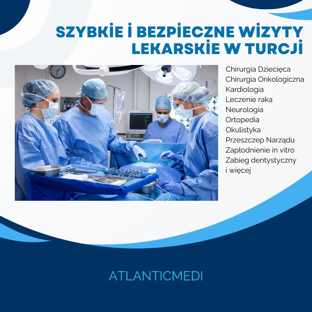
Türkiye can be the most convenient and fastest option.
Is your surgery date not in the near future? If you need to have surgery or treatment most efficiently for your health problem, Türkiye can be the most convenient and fastest option.
You can reach us 7/24 hours to get information about all treatments's process including terms as well as pricing.